


News
Top Stories

Do mood fluctuations impact confidence in decision-making?
Study reveals that in the healthy adult population, fluctuations of mood do not interfere with confidence in decision-making.

Relationship between sleep bruxism, insomnia and anxiety
Researchers concluded that, although sleep bruxism has no direct association with insomnia, anxiety may act as a bridging factor between these complaints.

Can IQ and socioeconomic status interfere with children's reading fluency?
Researchers found that Intelligence Quotient (IQ) plays no role with reading fluency deficits in children, unlike socioeconomic status.
News

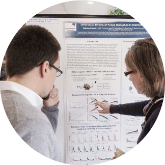
How does postingestive reward shape our choices?
Postingestive processes, which refer to the physiological and behavioural responses occurring after food intake, are crucial for regulating eating behaviour, influencing both the sensation of satiety and food preferences. The research team led by Albino Oliveira Maia investigated the impact of these processes on three distinct groups: healthy individuals, people with obesity, and post-bariatric surgery patients. Specifically, the study explored whether these effects occur through implicit (non-conscious) mechanisms and how the availability of dopamine D2 receptors in the brain is related to these processes. Using the flavour-nutrient conditioning (FNC) method, where the flavour of yogurt enriched with maltodextrin was associated with increased caloric content, it was observed that food preferences were influenced primarily through implicit consumption decisions rather than explicit assessments of flavour pleasantness ratings. In other words, the study demonstrated that FNC affected food choices without participants being consciously aware that they were preferring foods due to their association with nutritional value. This shift was observed in healthy individuals, people with obesity, and post-bariatric patients. Furthermore, people with obesity showed lower availability of dopamine D2 receptors, crucial in the brain's reward system. After bariatric surgery, particularly gastric bypass, there was an increase in receptor availability, which may be linked to improved feeding behavior regulation. These findings suggest that changes in the reward system may play a role in the surgery's effectiveness in reducing weight and modifying eating behaviours. This study was supported by the BIAL Foundation, in the scope of the research project 176/10 - Dopaminergic regulation of dietary learning in humans and rodents, and published in the journal PLOS BIOLOGY, in the article Postingestive reward acts through behavioral reinforcement and is conserved in obesity and after bariatric surgery.
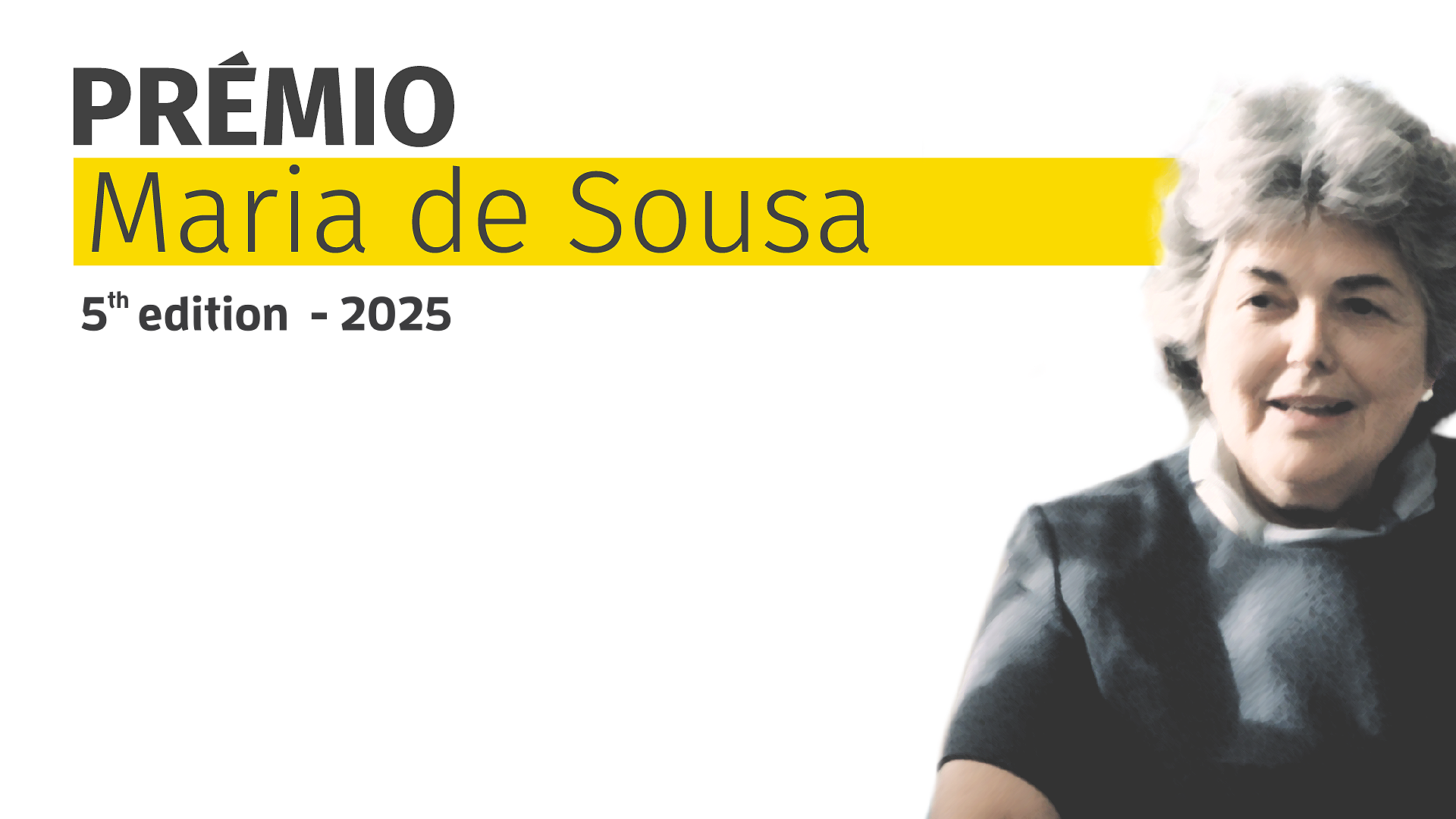
Maria de Sousa Award 5th edition - 2025: applications are open
Applications are now open for the Maria de Sousa Award 5th edition - 2025, promoted by the Portuguese Medical Association and the BIAL Foundation to honour the physician and great researcher Maria de Sousa. The deadline for applications is 31 May.

The BIAL Foundation has received 432 applications for the Grants Programme for Scientific Research
Did you know that the BIAL Foundation received 432 applications from 29 countries for the Grants Programme for Scientific Research 2024/2025, and 80 were approved?